
iStockphoto.com 179007773
In this follow-up to “Growth-plate injuries: A diagnostic challenge,” (May, page 15) we continue our look at physeal trauma, exploring growth-plate issues in anterior cruciate ligament reconstruction, and further examining these injuries’ impact on the knees, ankles, and feet of youth athletes.
By Shalmali Pal
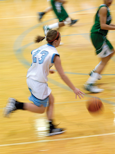
Surgeons are more likely to repair an injured anterior cruciate ligament (ACL) in young athletes than ever before, with studies showing the rate of reconstructions in skeletally immature knees tripling between 1990 and 2009.1-4
“ACL reconstruction [ACLR] in children is an important intervention,” said Mark V. Paterno, PT, PhD, MBA, SCS, ATC, coordinator of Orthopaedic and Sports Physical Therapy in the Division of Occupational Therapy and Physical Therapy at Cincinnati Children’s Hospital Medical Center in Ohio. “With the increased incidence of ACL injury in skeletally immature patients, a reliable option to restore mechanical stability in the knee is needed. Previous data have demonstrated that continued giving way or functional instability places patients at risk for further meniscal and articular cartilage injury.5 Ultimately, this can lead to an early onset of osteoarthritis. If children suffer an ACL injury and continue to have giving way in their knee, they need to have some option to increase its stability.”
ACLR can disturb growth plates, which typically close when boys are aged 15.6 to 17.1 years and when girls are aged between 15 and 16.9 years.5 Possible complications of ACLR in skeletally immature patients include premature growth arrest, angular deformity, limb overgrowth, and leg length discrepancy.6 Children with 5 cm or more of future lower limb growth potential are at greatest risk of iatrogenic growth disturbance caused by violation of the growth plate, according to a 2016 magnetic resonance imaging (MRI)-based analysis of the tibial epiphysis in skeletally immature adolescents.6
Physeal-sparing ACLR techniques, typically all-epiphysis techniques in which graft tunnels are drilled to align anatomically and to avoid growth-plate disruption,5 are gaining acceptance as a surgical option for complete ACL tears in skeletally immature knees (Figure 1). Yet treating ACL injuries without adversely impacting the open physis can be a challenge even with these techniques.6-8
Nawabi et al, for example, studied 15 skeletally immature knees after all-epiphyseal ACLR on quantitative MRI 24 months postoperatively. Ten of the 15 knees demonstrated violation of the tibial physis, typically at the anteromedial region where tunnel drilling occurred. The group’s mean volume of physeal damage was 2.1%, but two knees had a physeal injury greater than 6%.7 A 2017 meta-analysis of outcomes and complications of different ACLR techniques found the overall rate of growth disturbance after ACLR was 2.6%, with no significant difference between transphyseal and physeal-sparing techniques, though the latter had a lower rate of postoperative complications compared with transphyseal techniques.8
Paterno noted that while this research shows ACLR poses some risk to the physis, that risk is relatively low.
“Physeal-sparing ACL reconstruction may be a good option to restore stability at reduced risk to an open physis,” he said. “Although no procedure can eliminate risk, the physeal-sparing procedures may reduce risk of altered growth secondary to potential physeal injury while providing necessary stability in some patients.”
Nonsurgical treatment options are suitable for nondisplaced ACL injuries, such as Type 1 tibial eminence fractures, per the Modified Meyers and McKeever Classification.9 Treatment generally consists of three to four weeks of immobilization, either with long leg casting or splinting in extension.
Bracing after ACLR, preventive use
As for the role of bracing in ACL injuries, it’s often recommended after reconstruction with an eye toward controlling range of motion and protecting the graft as it heals.10 But there’s a fair amount of uncertainty about the value of knee bracing in this setting, such as whether it can mechanically protect a reconstructed ACL, and if long-term bracing will impact joint laxity and functional outcomes (see “Postop bracing after ACL reconstruction,” LER, January 2011, page 43).
“Bracing has not consistently been shown to reduce noncontact ACL injury rates. Braces are used inconsistently after ACLR, but, again, there is little evidence to confirm a reduced second injury rate in this population,” said Paterno. “Bracing may help improve proprioceptive awareness and protect against a contact injury in this population, which may be sufficient justification to support its use, as it may help increase patient comfort when they return to sports.”
Then there is the issue of using braces prophylactically to prevent injuries. Brian G. Pietrosimone, PhD, ATC, and colleagues conducted a systematic review of prophylactic braces for the prevention of knee ligament injuries in collegiate football players.11

Figure 1. Illustration of the knee depicting an all-epiphyseal ACLR. The arrow indicates the location of the entrance of the tibial tunnel at the anteromedial margin of the tibial epiphysis. (Reprinted with permission from reference 6.)
Some studies showed a relative risk reduction, while others actually indicated an increased risk of injury. But overall, the flawed methodology of the studies reviewed was enough for Pietrosimone’s group to conclude that “due to the inconsistent findings within the literature, we deem the current evidence regarding the efficacy of prophylactic knee bracing in reducing knee injuries inconclusive.”
The participants in the studies reviewed were young adults, but the efficacy of prophylactic bracing in children who are still growing is just as uncertain, said Pietrosimone.
“We wrote that [review] almost ten years ago, and the data that was part of that study was older than that; from the nineties,” said Pietrosimone, an assistant professor in the Department of Exercise and Sports Science at the University of North Carolina at Chapel Hill. “The braces that were studied in that paper are probably obsolete. Nobody has done what’s really needed—a randomized controlled trial to see if [prophylactic] braces are effective. But people are using them for their theoretical benefits.”
In 2001, however, the American Academy of Pediatrics (AAP) issued a technical report on the use of knee braces in young athletes, which was updated in 2015.12,13 The report acknowledged: “There is a lack of scientific evidence that these braces are helpful at the level required for athletic participation. However, patients report a positive subjective response, claiming an increase in knee stability, pain attenuation, performance enhancement, and confidence during athletics with brace use. … Brace wearers have higher energy expenditures than do nonwearers. Current experimental evidence suggests that [prophylactic] knee braces do not significantly affect performance.”
For Pietrosimone, the take-home message for practitioners about prophylactic bracing in this patient population is to proceed with caution. “I think it really comes down to the previous medical history of that athlete,” he said. “If you are talking about using bracing to prevent an injury in a person who has never had one, there’s not enough evidence to suggest this is a worthwhile use of your resources.”
He pointed out the AAP technical report covers a wide variety of knee braces—neoprene sleeves, padded knees braces, rigid braces—and that “these are all very different, and aren’t necessarily suitable across the board.”
For instance, a padded knee brace might make sense for a volleyball player, whereas a rigid collateral or rotational brace may not, given the player’s movement patterns.
“In the AAP technical report, some of the indications are for chronic knee issues … like trying to align the patella,” he noted. “A neoprene sleeve, in general, may be helpful for someone that has some sort of stiffness or chronic swelling; a sleeve may be useful to keep blood flow to the area. It could also provide some compression to the joint. But it may not necessarily improve stability.”
Knees, feet, and ankles
Physeal knee, foot, and ankle injuries can be put into three general categories: growth-related, overuse injuries, and acute injuries.
Growth-related injuries. The most common growth-related injury seen in young athletes is Sever disease, or an apophysitis of the os calcis. In young athletes, heel pain can be a tell-tale sign of an unfused apophysis. Sever disease is bilateral in the majority of cases, and affects male athletes more often.14 (See “Sever disease: Intervene early to relieve symptoms,” May 2015, page 15.)
Issues to look for during the physical exam are “posterior calcaneal tenderness with mediolateral compression anterior to the Achilles tendon insertion. Ankle dorsiflexion may aggravate the pain due to tight heel cords,” according to a Sports Health review article. Sever disease is often tied to forefoot pronation.14
“The differential diagnosis for calcaneal apophysitis includes tarsal coalition, osteomyelitis, retrocalcaneal bursitis, and neurologic disorders,” wrote the review authors.
Two good examples of coalition injuries in the foot are a talar calcaneal bar or a calcaneal navicular bar, according to Robert M. Conenello, DPM, a podiatrist with Orangetown Podiatry in Orangeburg, NY, and a past president of the American Academy of Podiatric Sports Medicine.
In young athletes, excessive pronation while running may increase the chances that they are setting themselves up for these types of injuries.15 He noted a young athlete may sustain these physeal injuries before they experience obvious symptoms. “When these areas start to ossify, they become more painful under stress … the patient may not notice there’s a problem until their midteens, when they start to add more bulk, strength, and weight,” said Conenello, who is also a clinical adviser for Special Olympics Fit Feet program. (see “Taller, heavier children have heightened Sever disease risk,” August 2015, page 5.)
He also described treating accessory ossicles in young athletes. Accessory ossicles are separate ossification centers located extrachondrally, and they are different from coalitions because they do not form a connection between two bones but exist at the end of certain bones.16
Accessory ossicles generally appear when children are aged 8 to 10 years, and fuse about a year after their formation. According to a review article, “When they do not fuse, they become symptomatic. The most common sites for accessory ossification center formation are at the posterior talus, known as os trigonum, the medial malleolus, and the navicular. The navicular ossification center sometimes can form an entirely new bone known as an accessory navicular.”16
As with other physeal injuries, accessory ossicles can be misdiagnosed in the urgent care or emergency department (ED) setting. “I had this happen … with one of the kids on my son’s baseball team,” Conenello explained. “He said they’d gone to the [ED] and they told him he had a fracture. I asked to see the x-ray and said, ‘That’s not a fracture; that’s an accessory bone.’ I asked if they took an x-ray of the other foot because sometimes you’ll see the same problem on the other side.”
Overuse injuries. Then there are the overuse injuries, such as tibial tubercle apophysitis (Osgood-Schlatter disease) and osteochondroses. Osgood-Schlatter disease is often seen with activities that call for repeated forced knee extension. This extensor mechanism causes repetitive tensile microtrauma at the tibial tubercle apophysis.14
“The patellar tendon inserts at the unossified distal portion of the tibial tubercle apophysis via fibrocartilage. When this weak secondary ossification center is unable to withstand the repetitive tensile forces, bony or cartilaginous separation occurs, usually during the preossification or ossification phase; bone formation between the fragments follows,” the review authors wrote.
iStockphoto.com 172146706
It’s more common among boys and can be bilateral in more than half of the cases. The symptoms—gradual onset of pain, swelling, tenderness, and/or a prominent at the tibial tuberosity region—can worsen with jumping or running.16
Osteochondroses refer to lesions of the ossification centers that eventually undergo recalcification. The two most common lesions are osteochondrosis of the tarsal navicular (Kohler disease) and osteochondrosis of the second or third metatarsal heads (Freiberg infarction). These injuries can occur with up to 6.5% of ankle sprains and present as a chronic condition associated with an acute injury.16
The last of the overuse injuries are stress fractures, described as a process that leads to fatigue or insufficiency or failure of bone that occurs when the bone’s reparative abilities have been surpassed and the bone is unable to withstand chronic repetitive submaximal loads. These injuries account for up to 15% of all athletic injuries in young athletes, and are commonly seen in adolescent runners, but can occur during any sport that requires repetitive running or cutting actions.15
Acute injuries. Finally, there are acute physeal acute fractures, which are covered more extensively in part one of this series. Generally classified with the Salter-Harris system, acute fractures of the foot and ankle are most commonly are seen in the distal tibia, distal fibula, and the phalanges.
While the experts in part one discussed problems with misdiagnosis of these fractures, Conenello brought up another concern, that of overtreatment.
“I think misdiagnosis can go both ways,” he said. “Physeal fractures may be missed in EDs and trauma centers. But, sometimes, they’ll call it correctly as a growth plate fracture, and [subsequent physicians] will overtreat it. So, a patient will be in a CAM boot walker for six weeks. Then they get out of the boot and someone says to the kid, ‘Now get back on that soccer field.’ Now the athlete is deconditioned, and is set up for another potential injury down the line.”
And that dovetails with Conenello’s general philosophy when it comes to treating physeal injuries of the foot and ankle—he encourages his patients to work on their strength and balance.
“The more important thing to teach is strength because a strong athlete is one who is less likely to sustain injuries,” he noted. “As parents … we drive them from practice to practice [where] they work super hard. But in between practices, we don’t necessarily work on things like balance, strength, figuring out their weaknesses and what they could work on.”
For example, Conenello said he has treated gymnasts with Sever disease. During the exam, he said he’ll ask them to stand and balance on one foot. “And they fall—they’re gymnasts! Sure, they can do back walkovers and jump up in the air and twist around, but they aren’t strong enough yet. I’ll tell them to brush their teeth while standing on one foot, and they struggle with it. It’s overuse coupled with a certain lack of strength, especially in their feet and ankles.”
While he may use an over-the-counter (OTC) insole to address the heel pain in the acute phase, he is not apt to use devices long term. Instead, he will send patients for preferred body-weight training or plyometrics as ways to gain strength and mobility. “I think balance and proprioception are important in terms of being able to feel where their bodies are in space,” he explained.
Another issue Conenello will address in patients with physeal injuries is footwear, both on and off the playing field, court, or pitch. He noted kids will often gravitate toward athletic shoes because of the style, or because they are endorsed or designed by their favorite sports star. But that particular shoe may be inappropriate in terms how much flexibility it provides, how rigid it is, or the amount of room in the toe box.
“You have to really look at how the shoe fits on that kid,” he said. “I like shoes with wide toe boxes and some flexibility in the outsole. You also have to ensure that the kid is wearing the shoes properly.”
For instance, a child in basketball shoes who complains of heel pain may be tying the laces loosely, which is fashionable but not supportive of their feet and ankles (See “Basketball shoes trends favor fashion over feet, LER, February 2017, page 43, and “Seeking shoe closure: Laces versus alternatives,” LER Foot Health, October 2015, page 15).
Finally, Conenello said he always asks about shoe choices when the young athlete is not at play. He shared the story of a 13-year-old gymnast who complained of foot pain from a physeal injury, and when he asked her about her everyday footwear, she replied that she wore either flip flops, sheepskin-lined boots, or lace-up sneakers with a very flat, rigid insole.
“I told her: ‘That’s the problem. You spend the majority of your day in shoes that aren’t giving you the right support.’ I prescribed an OTC insole to educate her about what a supportive shoe should feel like. It’s important to teach good foot health outside of the sport.”
Shalmali Pal is a freelance writer in Tucson, AZ.
- Dodwell ER, Lamont LE, Green DW, et al. 20 years of pediatric anterior cruciate ligament reconstruction in New York State. Am J Sports Med 2014;42(3):675-680.
- Kocher MS, Saxon HS, Hovis WD, Hawkins RJ. Management and complications of anterior cruciate ligament injuries in skeletally immature patients: survey of the Herodicus Society and The ACL Study Group. J Pediatr Orthop 2002;22(4):452-457.
- McCarroll JR, Shelbourne KD, Porter DA, et al. Patellar tendon graft reconstruction for midsubstance anterior cruciate ligament rupture in junior high school athletes. An algorithm for management. Am J Sports Med 1994;22(4):478-484.
- Stanitski CL, Harvell JC, Fu F. Observations on acute knee hemarthrosis in children and adolescents. J Pediatr Orthop 1993;13(4):506-510.
- Dunn KL, Lam KC, Valovich McLeod TC. Early operative versus delayed or nonoperative treatment of anterior cruciate ligament injuries in pediatric patients. J Athl Train 2016;51(5):425-427.
- Davis DL, Almardawi R, Mitchell JW. Analysis of the tibial epiphysis in the skeletally immature knee using magnetic resonance imaging: an update of anatomic parameters pertinent to physeal-sparing anterior cruciate ligament reconstruction. Orthop J Sports Med 2016;4(6):2325967116655313.
- Nawabi DH, Jones KJ, Lurie B, et al. All-inside, physeal-sparing anterior cruciate ligament reconstruction does not significantly compromise the physis in skeletally immature athletes: a postoperative physeal magnetic resonance imaging analysis. Am J Sports Med 2014;42(12):2933-2940.
- Longo UG, Ciuffreda M, Casciaro C, et al. Anterior cruciate ligament reconstruction in skeletally immature patients : a systematic review. Bone Joint J 2017;99-B(8):1053-1060.
- Mall NA, Paletta GA. Pediatric ACL injuries: evaluation and management. Curr Rev Musculoskelet Med 2013;6(2):132-140.
- Little RM, Milewski MD. Physeal fractures about the knee. Curr Rev Musculoskelet Med 2016; 9(4):478-486.
- Pietrosimone BG, Grindstaff TL, Linens SW, et al. A systematic review of prophylactic braces in the prevention of knee ligament injuries in collegiate football players. J Athl Train 2008;43(4):409-415.
- Martin TJ, the Committee on Sports Medicine and Fitness. Technical report: knee brace use in the young athlete. Pediatrics 2001;108(2):503-507.
- AAP Council on Sports Medicine and Fitness. Knee Pain: How to Choose the Right Knee Brace for Your Child. Healthychildren.org. https://www.healthychildren.org/English/health-issues/injuries-emergencies/sports-injuries/Pages/Knee-Pain-and-braces.aspx. Updated November 21, 2015. Accessed August 10, 2017.
- Frush TJ, Lindenfeld TN. Peri-epiphyseal and overuse injuries in adolescent athletes. Sports Health 2009;1(3):201-211.
- Houghton KN. Review for the generalist: evaluation of pediatric foot and ankle pain Pediatr Rheumatol Online J 2008;9;6:6.
- Malanga GA, Ramirez-Del Toro JA. Common injuries of the foot and ankle in the child and adolescent athlete. Phys Med Rehabil Clin N Am 2008;19(2):347-371.
Related Posts
 October 24, 2012 Juvenile idiopathic arthritis study reveals footcare information gap
 October 3, 2022 DAJTEMU
 February 29, 2016 Keeping kids in braces can prevent clubfoot relapse
 October 2, 2022 DAJTEMU
 April 5, 2018 Media, toys, and games for kids with disabilities